Hemianopia: Types, Causes, Treatment and More
Hemianopia is an eye condition that describes losing half of your field of vision because of damage to the optic nerve in the brain.
Sight loss can occur in either eye or it can occur in both eyes at once.
Among the causes are diagnosed cases of multiple sclerosis (MS), Alzheimer’s disease, epilepsy and dementia.
Chemotherapy and low vision therapy are two types of treatments, but treatments vary based on the cause of the sight loss.
Intro
Hemianopia, also known as hemianopsia, is a condition in which you lose half of your vision field. Vision loss can be to the right side or the left side, and multiple types of brain damage cause it.
The condition can be temporary or permanent, depending on the cause.
Types of Hemianopia
There are several types of hemianopia, depending on the part of the eye it affects and the extent to which you lose vision. These types are:
- Homonymous
- Heteronymous
- Superior
- Inferior
- Homonymous quadrantanopia
Homonymous
Homonymous hemianopia occurs when you lose half of your vision field on the same side of each eye. Usually the side of brain damage is opposite to the side of vision loss. If brain damage occurs on the right side, vision loss will be on the left side.
Heteronymous
Heteronymous hemianopia is when you lose half of your vision field on different sides of your eyes. It can either be:
- Bitemporal, where you lose sight on the outer halves of each eye; or
- Binasal, where you lose sight on the inner halves of each eye
Superior
This is when you lose vision of the upper half of each eye.
Inferior
Inferior hemianopia is when you lose sight of the lower half of each eye.
Homonymous Quadrantanopia
This occurs when you lose a quarter of your vision in each eye.
Partial vs. Complete Hemianopia
Partial and complete hemianopia differ, depending on how much vision field is lost. Partial hemianopia occurs when a quarter (quadrant) of your vision field loses sight.
But for complete hemianopia, the loss of sight occurs in half of your vision field. It’s the role of the National Institute of Health Stroke Scale (NIHSS) to determine whether hemianopia is partial or complete.
Causes of Hemianopia
The leading cause of hemianopia is stroke. But hemianopia can result from anything that causes damage to the optic nerve of the brain. These causes included a tumor and traumatic injury to the brain. Other causes include:
- Multiple sclerosis
- Alzheimer’s disease
- Epilepsy
- Dementia
- Neurosyphilis
- Lymphoma
- Shaken baby syndrome
- Neurosurgical procedures
- Abnormal blood vessels
- Hydrocephalus
- High pressure in the brain
Symptoms
It might not be easy to tell that you have hemianopia. You might feel like you have a problem with your right eye if you can’t see the right side well. If you have left hemianopia, you might think your left eye has a problem.
Primary symptoms of hemianopia are:
- Failing to see objects on the affected side, such as lanes on the road and food on the table
- Loss of color vision
- Double vision
- Visual hallucinations. It involves seeing shapes and light flashes
- Losing depth perception in case of heteronymous hemianopia
- Bumping into things because you can’t see them
- Difficulty with spatial relationships (hard to judge the space between you and another object)
- Difficulties maneuvering crowded streets
- When walking, you might drift away from the impaired side
- Failure to see the entire block of text when reading
- Lower night vision
- Having a dimmed vision
- Failure to understand what you see
- Distorted sight or blurry vision
Diagnosis
Even though hemianopia results from brain damage, doctors diagnose the condition by examining the eyes. Your doctor will perform a visual field test to determine the size of the affected area and the location.
The doctor will also examine the inside parts of your eyes to rule out the possibility of a different cause.
In addition to examining your eyes, your doctor may require you to get an MRI (magnetic resonance imaging) to check your brain’s health.
Treatment
Doctors build treatment regimens for hemianopia based on the cause of each case. If caused by a tumor, surgery and chemotherapy can help cure the problem. In case of stroke and head injuries, you might regain vision with time.
If your sight fails to improve, a low vision specialist can help you make the best use of the remaining sight. That typically means low vision therapy.
Low vision therapy requires you to attend a rehabilitation program to learn how to do daily activities primarily on your own. These activities include grooming, preparing food, self-care, reading, navigating your environment and more.
Other strategies to help you cope with the problem include:
- Use of visual aids to help you see the missing fields
- Learning to turn your body or head to the impacted side to get a broader view
- Having a walking partner to guide you
- Placing your hand on the edge of the page to help you read the full text
- Using a straight edge to point to the line you’re reading
- Scanning your surroundings to know the location of people and objects
- Adjusting your sitting position to get a better view of people during a conversation or when watching the television
- Playing puzzle games and crosswords to gain eye coordination
- Using a driving simulator to determine your ability to drive safely
Outcomes
You sometimes can recover from hemianopia without treatment. If the no-treatment option does not work, you must receive treatment for the underlying cause to regain full sight.
If the problem is untreatable, your hope is low vision therapy. With successful rehabilitation, you might be able to improve on activities like reading, navigating your environment, perhaps even driving.
FAQs
Is hemianopia the same as hemianopsia?
Hemianopia is the same as hemianopsia. Both terms mean losing half of the visual field due to damage to the brain.
What is hemianopia in stroke?
Hemianopia in stroke is a loss of half of the visual field that results when a person gets a stroke. Effects on the eyes depend on the extent and location of the stroke on the brain. If a stroke occurs on the right side of the brain, vision impairment results on the left side of the eyes and vice versa.
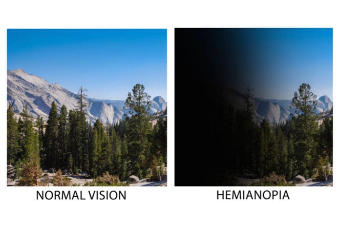
What does hemianopia look like?
Hemianopia leads to a narrowed view and a distorted sight. You may stumble on objects because you can’t see them. Plus, you might have challenges walking straight and reading entire blocks of text.
References
-
Homonymous Hemianopsia. (April 2021). Cleveland Clinic.
-
Stroke-related Vision Loss. Johns Hopkins Medicine.
-
Chapter 12 – reading and alexia. (2021). Handbook of Clinical Neurology.
-
An Overview of Hemianopia. (May 2020). Very Well Health.
-
Chapter 14 – Unilateral spatial neglect after posterior parietal damage. (2018). Handbook of Clinical Neurology.
-
Chapter 10 – Rehabilitative techniques. (2011). Handbook of Clinical Neurology.
Last Updated April 4, 2022
Note: This page should not serve as a substitute for professional medical advice from a doctor or specialist. Please review our about page for more information.