Retinal Dystrophies: Causes, Symptoms and Treatments
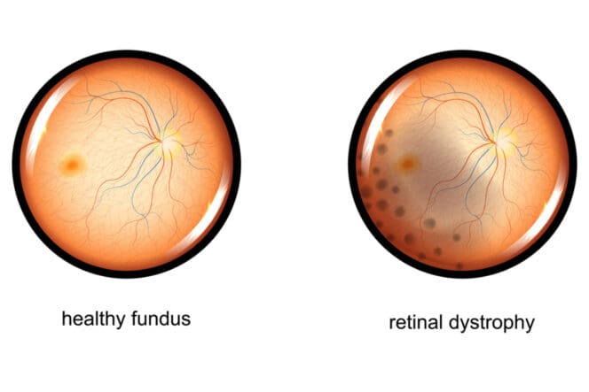
Retinal dystrophies are hereditary illnesses that strike the retina and choroid, causing progressive and severe vision loss. None of them have cures.
Among the conditions they cause include retinal tears, retinal detachments, macular degeneration and retinitis pigmentosa.
Symptoms include night blindness, altered color perception and photophobia.
What Is Retinal Dystrophy?
The retina is one of the integral parts of the eye. Damage to it can lead to devastating effects, including vision loss. There are a certain group of genetic diseases that can affect the choroid and retina. These diseases are known as retinal dystrophies.
Retinal dystrophies are various types of hereditary illnesses that cause severe and progressive vision loss. The diseases alter the functioning and anatomy of the retina, causing an array of complications.
Medical science has no known cure, and the diseases’ low prevalence rate doesn’t help with the extensive research needed to find curative measures.
Retinal dystrophies can affect the photoreceptor cells, either rods or cones. In some cases, diseases strike all photoreceptor cells.
Among the retinal dystrophy diseases that affect both the rods and cons are retinitis pigmentosa and Stargardt disease.
Diseases Classified as Retinal Dystrophies
The more common retinal dystrophy diseases and conditions include:
- Diabetic retinopathy
- Epiretinal membrane
- Macular degeneration
- Macular hole
- Retinal detachment
- Retinal tear
- Retinitis pigmentosa
Diabetic Retinopathy
If you have diabetes, the capillaries at the back of your eye can deteriorate and leak fluid into and around your retina. When this happens, the retina can swell, leaving you with blurry and distorted vision.
In some cases, someone with diabetes develops new, abnormal capillaries that break easily and bleed. The more bleeding that occurs, the more vision is affected.
Epiretinal Membrane
The epiretinal membrane is a fragile tissue-like scar that looks like crinkled cellophane atop the retina. When this membrane pulls up on the retina, it can affect your vision and make objects appear crooked or blurry.
Macular Degeneration
People with macular degeneration can experience deterioration of the retina from the center. This deterioration can lead to further symptoms, including blurred central vision or the formation of a blind spot in the center of your field of vision.
There are two types of macular degeneration: dry and wet macular degeneration. Most people will first develop dry macular degeneration before it progresses to wet macular degeneration in one or both eyes.
Macular Hole
This is a condition in which a small defect, or hole, forms at the center of the retina and back of the eye (or macula). The hole can come from an injury to the eyes or from abnormal movement between the vitreous and retina.
Retinal Tear
Retinal tears occur when the vitreous shrinks and pulls on a thin layer of tissue lining the retina. Continuous tugging breaks the tissue. The most noticeable symptoms include sudden vision changes, including flashing lights and floaters.
Retinal Detachment
This condition manifests when fluid resides under the retina and passes through a tear in the retina. Fluid causes the retina to lift from its place, leading to various abnormalities.
Retinitis Pigmentosa
Retinitis pigmentosa is a hereditary degenerative disease that gradually affects the retina and may cause loss of vision at night and side vision.
Causes of Retinal Dystrophy
Retinal dystrophies are genetic and get passed down from one generation to the next. They exhibit different types of inheritance patterns about who in younger generations receives the genetic code to a specific malady.
The most prevalent inheritance patterns are the dominant autosomal, recessive autosomal and the X-chromosome-linked inheritance.
Dominant Autosomal
The gene affects all generations in an affected family. In dominant inheritance, all blood family members carry the mutation and eventually transmit it to approximately half of all descendants. An example of retinal dystrophies passed through dominant inheritance is the Best disease, or Best vitelliform macular dystrophy.
Recessive Autosomal
Children whose parents are carriers have a 25 percent chance of having recessive autosomal. The probability of development remains constant during each pregnancy and affects both males and females equally. An example of retinal dystrophies passed through recessive autosomal is Stargardt disease.
X Chromosome-Linked Inheritance
In these cases, only men in a family suffer from the illness, although the women are also carriers. Because women carry the gene, they have a 50 percent chance of passing the disease to male children. An example of retinal dystrophy passed through the X link chromosome is choroideremia.
Symptoms of Retinal Dystrophy
Retinal dystrophy’s speed of evolution and symptoms depend on the person and the type of retinal dystrophy disease they have. But some common signs and symptoms of retinal dystrophies exist. Among them are:
- Night blindness: This is the inability to properly adapt your sight in dimly lit places. Night blindness can greatly affect your normal life, especially during the night, as you’ll have a challenge discerning different objects, movements, and characteristics.
- Altered color perception: You may find it difficult to discern among different colors. This is an especially worrying symptom as it usually occurs when the disease has progressed to advanced stages.
- Visual field changes: Some retinal dystrophies cause the peripheral visual field to contract. However, the central vision can remain intact until further stages of the disease. In other cases, the disease can solely affect the central visual field and reduce your visual acuity.
- Metamorphopsia: This is the presence of a wave-like appearance on straight lines. This characteristically reveals significant changes in the macula.
- Photophobia and glare: Photophobia is a discomfort caused by flickering or flashing lights in high-light conditions. Moving from bright or dark environments or vice versa can also cause photophobia.
Retinal Dystrophy Treatment
Part of the difficulty in treating retinal dystrophy is the challenge of regenerating affected retina cells. However, there are a few promising treatment options being extensively studied.
These prospective remedies include the designing and applying of cell and gene therapies. These therapies might be able to restore or slow down the progression of vision loss.
Diagnosing Retinal Dystrophies
While there are varying methods of diagnosing retinal dystrophy, the most common is a direct examination of the retina. In some cases, complementary tests may be used to identify the exact type of retinal dystrophy disease.
These complimentary tests may include autofluorescence exams and optical coherence tomography. A genetic test may also be required to determine the presence of mutated genes contributing to the disease’s progression.
Coping with Vision Loss
The sense of sight is arguably the most relied upon. Losing your vision greatly affects your mental and emotional health.
Eye professionals advise seeking counseling services if you are facing mental and emotional problems from vision deterioration. In addition, the following tips can help you cope better with vision loss:
- Learning more about the condition
- Seeking therapeutic counseling services
- Understanding the grieving and adjustment periods
- Exploring the benefits of adjustment devices and classes
Low Vision Aids for Retinal Dystrophy
Numerous low-vision aids and non-optical devices can be modified to help those with lowered vision. These aids and devices include:
- Reading prisms
- Hand magnifiers
- Telescopic glasses
- Magnifying glasses
- Light-filtering lenses
- Closed-circuit television
Computer software also exists to alter certain images to make them more clear and also and read types of screen text.
References
-
Age-related Macular Degeneration. (June 2021). National Eye Institute.
-
Vitreous Detachment. (September 2020). National Eye Institute.
-
Retinal Dystrophies and the Road to Treatment: Clinical Requirements and Considerations. (June 2020) National Center for Biotechnology Information.
-
Macular Edema. (March 2018). American Society of Retina Specialists.
-
Prevalence of Generalized Retinal Dystrophy in Denmark. (August 2014). National Center for Biotechnology Information.
Last Updated April 20, 2022
Note: This page should not serve as a substitute for professional medical advice from a doctor or specialist. Please review our about page for more information.