Central Heterochromia: Types and Causes
Central heterochromia describes an uncommon condition in which the inner and outer rings of the iris are different colors.
Most people who have central heterochromia get it from a family history, but there are sporadic cases of non-genetic causation.
The condition is linked to a small variety of illnesses but is usually not serious.
What Is Central Heterochromia?
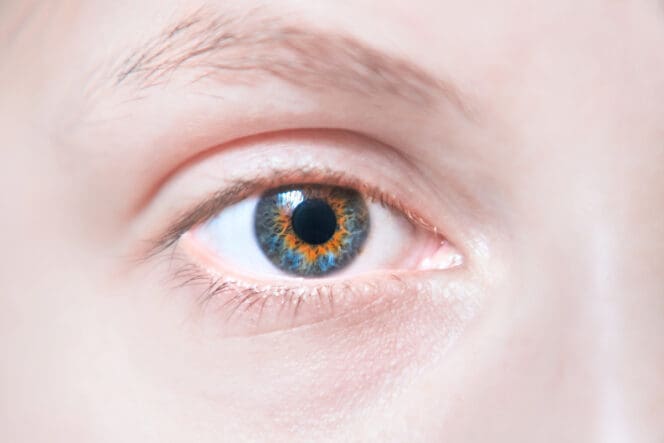
Central heterochromia is a condition in which the inner ring of your iris (the section closest to your pupil) has a different coloration from the outer ring of your iris (area along the edge of your iris). This condition usually affects both eyes.
The exact prevalence of central heterochromia is not known since the condition itself rarely requires medical attention and is thus difficult to document. In the United States, fewer than 200,000 people are affected by complete heterochromia, where the color of one iris is different from the other. Central Heterochromia is known to be more common than complete Heterochromia.
Causes
Most cases of central heterochromia stem from genetics and are present from birth. When this is the case, the condition is called congenital heterochromia. Central heterochromia can occur without any underlying abnormality and is typically benign.
According to the Genetic and Rare Diseases Information Center, many heterochromia cases occur sporadically in individuals with no familial history of the eye condition. Some cases of congenital heterochromia are linked to the following syndromes and diseases:
- Horner’s syndrome
- Bloch-Sulzberger syndrome
- Bourneville disease
- Sturge-Weber syndrome
- Hirschsprung disease
- Parry-Romberg syndrome
- Waardenburg syndrome
- Von Recklinghausen disease
Other cases of central heterochromia develop later in life, triggered by an injury, illness or medication. The condition is then termed acquired heterochromia and is less common than the genetic type. Acquired heterochromia can be caused by:
- Injury to your eye
- Glaucoma
- Diabetes mellitus
- Acquired Horner’s syndrome
- Iris ectropion syndrome
- Eye surgery
- Pigment dispersion syndrome
- Iris tumors
- Posner-Schlossman syndrome
- Iris tumors
- Eye swelling
Also, a medication used to treat glaucoma, latanoprost, may cause heterochromia. Latanoprost is associated with iris color changes in some patients undergoing long-term latanoprost therapy.
How Is Central Heterochromia Different from Other Forms of Heterochromia?
Heterochromia refers to a variation in the colors of the iris. Central heterochromia is one of three primary main types of heterochromia that include complete heterochromia, or partial heterochromia.
In central heterochromia, an iris shows two colors. But the arrangement of the colors is concentric with the central zone of the iris being a separate color from the mid- to peripheral zone. Here is how it differs from complete and sectoral heterochromia:
Complete Heterochromia
Complete heterochromia is the condition where one iris has a different color than the other iris. For instance, one eye may be blue and the other eye may be brown. It is the rarest form of heterochromia.
Sectoral Heterochromia
This is also referred to as partial heterochromia. It is the condition where part of one iris has a different type of color from the remaining areas of that iris. It usually resembles an irregular patch on your iris and doesn’t form a ring around your pupil.
This eye condition can affect one or both eyes. Like the complete form, sectoral heterochromia may be acquired or associated with clinical syndromes. The exact prevalence of this condition is unknown, but it is less common in humans than complete heterochromia.
What Causes the Different Colors in Central Heterochromia?
Understanding the source of eye color variation is important to understand what causes the different colors in central heterochromia. The concentration and distribution of melanin primarily determine your eye color and, specifically, the color of your irises.
Researchers do not fully understand the processes that determine eye color, but multiple genes determine your inherited eye color. Acquired or environmental factors may alter these inherited characteristics.
There are two pigments present in your iris, eumelanin and pheomelanin. The concentration of these pigments, distribution variation of the pigments in the stroma layers in your iris, the ratio between the pigments, and light scattering effects play a role in determining your eye color.
Heterochromia is noticeable in terms of irises that contain low levels of melanin. A common form of the eye condition presents predominantly in blue eye, where a smaller ring of brown or gold streaks outwards from the pupil.
The color variation in the iris of people with Heterochromia is due to the melanin pigments being distributed differently towards the central zone of the iris closer to the pupil. The definitive cause of this variation is unknown. Light reflects off the different pigmented sections of the iris in different ways. As a result, it gives the appearance of two separate colors in each iris.
Treatment
Heterochromia, especially the congenital form, is a typically harmless eye condition. In most cases, this condition does not cause any health complications or affect your vision. As such, treatment is usually not necessary if no other underlying problems are present.
However, if your central heterochromia is acquired, it is important to seek medical attention to address the underlying condition. You can use colored contact lenses if you want to change how your eyes look for cosmetic reasons.
References
-
Heterochromia. (August 2008). Canadian Medical Association Journal.
-
What is Heterochromia? (April 2021). American Academy of Ophthalmology.
-
Heterochromia iridis. (April 2015). Genetic and Rare Diseases Information Center.
-
Incidence of iris color change in latanoprost treated eyes. (October 2002). British Journal of Ophthalmology.
-
Characterization of Melanins in Human Irides and Cultured Uveal Melanocytes from Eyes of Different Colors (September 1998). Experimental Eye Research.
-
The color of the human eye: A review of morphologic correlates and of some conditions that affect iridial pigmentation. (February 1997). Survey of Ophthalmology.
Last Updated April 20, 2022
Note: This page should not serve as a substitute for professional medical advice from a doctor or specialist. Please review our about page for more information.