Open-angle Glaucoma: Risk Factors, Signs, Causes, and Treatment
Open-angle glaucoma occurs when an eye is unable to drain fluid as effectively as it should despite the drainage angle remaining open. With the fluid not draining fast enough, increased pressure inside the eye can damage the optic nerve.
The heightened pressure wreaks havoc slowly. First it causes a loss of one’s progressive peripheral vision, followed by loss of central vision.
The condition does not cause any pain, but it can cause irreversible blindness if left untreated. Most people with the condition do not experience symptoms until they start losing their vision.
The most common type of glaucoma, it accounts for about 80 percent of all glaucoma cases in the United States.

Symptoms
Early Symptoms
What makes open-angle glaucoma so difficult to treat effectively is that its early symptoms are so difficult to detect. With no discernable discomfort and no loss of vision from blurriness, eye floaters or other eyesight issues, people who develop this malady are mostly unaware until it is beyond the early stage.
That makes the condition asymptomatic until significant visual field changes occur.
Later Symptoms
Someone with open-angle glaucoma starts to show symptoms in later stages when they become aware of blurred vision or constricted visual field. Vision loss starts in your side or peripheral vision as the condition progresses.
You may find yourself turning your head sideways to compensate for the blind spots in your side vision. You may also learn of the early visual defects when carrying out monocular tasks like using a camera’s viewfinder. Peripheral vision loss in elderly patients may lead to difficulty doing tasks like driving and running into objects in the home more frequently.
Besides peripheral vision loss or reduced sight, other later-stage symptoms may include:
- Swollen or bulging cornea
- Red eye
- Nausea
Without effective control of the disease at the stage where it causes peripheral vision loss, it can lead to tunnel vision, limiting your field of vision to only images straight ahead or in your central vision. As the glaucoma progresses, central vision loss occurs as well, leading to complete or partial permanent vision loss.
Causes
Researchers are unsure of the exact causes of open-angle glaucoma. There is consensus that the cause is related to the obstruction of aqueous outflow through the trabecular meshwork rather than the production of too much aqueous.
Scientists have proposed several mechanisms for the obstruction, such as oxidative damage, a buildup of foreign material, an immunological process, or phagocytic activity loss.
The progressive resistance to aqueous outflow causes the gradual increase of the pressure inside the eye, or intraocular pressure (IOP), which could make someone susceptible to glaucomatous optic nerve damage.
Risk Factors
Genetic and other risk factors for developing open-angle glaucoma that scientists have studied and extensively described include:
- Elevated IOP
- Family history of glaucoma
- Older age
- African ancestry
- Systemic diseases like hypertension and diabetes
- Thinner cornea
- Low blood pressure
Diagnosis
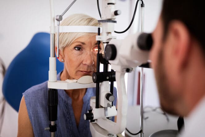
Eye doctors use several diagnostic tools for clinical evaluation of open-angle glaucoma. Among them:
- CCT measurement: Thin central corneal thickness (CCT) is a risk factor for developing open-angle glaucoma. Doctors measure CCT to help interpret IOP readings and stratify patients for ocular damage.
- Visual field testing: Visual field testing, also known as perimetry, maps out the visual field of patients on a printout. The test helps doctors and patients visualize exact sight loss. Over time, more visual field testing maps out the speed and magnitude of vision loss.
- Gonioscopy: Gonioscopy helps doctors rule out other ocular emergencies like closed-angle glaucoma. During this test, your eye doctor will numb your eyes and place a special contact lens prism on the eye surface to determine whether the angle is closed or open.
- OCT scan: An optical coherence tomography (OCT) scan produces high-resolution images of the optic nerve, anterior segment, and retina. The cross-sectional imaging of the anterior angle facilitates the detection of angle-closure or open-angle glaucoma.
Open-Angle vs. Closed-Angle Glaucoma
For both open-angle glaucoma and closed-angle glaucoma, increased intraocular pressure is a considerable risk factor. However, they also have significant differences:
- In open-angle glaucoma, the angle where the iris and cornea meet remains open as it is supposed to be, but in closed-angle glaucoma, the angle is narrow.
- Open-angle glaucoma develops slowly, but closed-angle glaucoma can develop quickly.
- Open-angle glaucoma has signs and damage that are usually not noticeable. Closed-angle glaucoma can have symptoms and damage that are easily noticed.
Treatments
Open-angle glaucoma usually responds well to treatment if detected early and treated. The aim of all treatment strategies is to lower eye pressure.
Eye specialists tend to begin treatment with topical or oral medications. In case of progressive damage, they may consider treatments like laser trabeculoplasty, trabeculectomy, or drainage implant.
Medication, surgery or a combination of the two often slows down the progression of open-angle glaucoma.
Glaucoma Medication
Eye drops can control many open-angle glaucoma cases. There are several types of eye drops that work differently to control intraocular pressure. For example, prostaglandins relax your eye muscles to facilitate better drainage of fluid and reduce IOP buildup. A variety of eye drops use beta-blockers, which decrease the amount of fluid produced.
In many cases, doctors give patients more than one type of anti-glaucoma drug.
Eye surgeons have three go-to procedures as a way to treat this type of glaucoma:
- Trabeculoplasty
- Trabeculectomy
- Glaucoma Drainage Implant
Trabeculoplasty
Doctors can consider laser trabeculoplasty as the initial therapy for some open-angle glaucoma patients or as an alternative for people who will not or cannot use medication because of intolerance to medication, memory problems, cost, or difficulty with instillation. During the procedure, doctors may use argon, diode, or frequency-doubled Nd:YAG (neodymium: yttrium-aluminum-garnet) lasers to improve the functioning of the drainage angle.
Laser trabeculoplasty improves aqueous outflow, helping lower IOP.
Trabeculectomy
This procedure involves a surgeon creating a tiny opening in the sclera under your eyelid to give aqueous humor an alternative path for draining out of your eye. The tissue around the eye absorbs the fluid, lowering eye pressure.
Doctors generally perform trabeculectomy when medication and laser therapy are not enough to control a patient’s open-angle glaucoma. In some cases, they can consider it as the initial therapy.
Glaucoma Drainage Implant
An ophthalmologist may implant a small drainage tube to send the fluid to a reservoir. The surgeon will create the reservoir beneath your conjunctiva. The nearby blood vessels will then absorb the fluid.
Prevention
Because open-angle glaucoma has few warning signs before severe damage occurs, the best way to avoid vision loss is by having regular eye examinations, especially if you are over 40 years old.
If your eye doctor detects the disease during an exam, the doctor may prescribe preventive treatment to help preserve your vision.
For instance, some people are at a higher risk of getting open-angle glaucoma due to a raised IOP. Data from the Ocular Hypertension Treatment Study (OHTS) and the European Glaucoma Prevention Study (EGPS) has shown that using topical therapy to reduce IOP can lower the risk of such people developing open-angle glaucoma.
References
-
Open Angle Glaucoma. (August 2021). StatPearls Publishing.
-
The Ocular Hypertension Treatment Study. (June 2002). JAMA Ophthalmology.
-
Primary Open-Angle Glaucoma in Blacks: A Review. (May 2003). Survey of Ophthalmology.
-
Genetic Risk of Primary Open-angle Glaucoma. (December 1998). JAMA Ophthalmology.
-
Q-switched 532-nm Nd:YAG laser trabeculoplasty (selective laser trabeculoplasty). (November 1998). American Academy of Ophthalmology.
-
Primary Open-Angle Glaucoma Preferred Practice Pattern Guidelines. (November 2015). American Academy of Ophthalmology.
-
What Is Glaucoma? Symptoms, Causes, Diagnosis, Treatment. (September 2021). American Academy of Ophthalmology.
-
Primary Open-Angle Glaucoma. (December 2021). American Academy of Ophthalmology.
-
Symptoms of Open-Angle Glaucoma. (December 2019). Glaucoma Research Foundation.
-
What Is Open Angle Glaucoma? (July 2020). Optometrists Network.
-
Glaucoma Facts and Stats . (October 2017). Glaucoma Research Foundation.
-
Glaucoma and Eye Pressure. (September 2021). National Eye Institute.
-
Types of Glaucoma. (September 2021). National Eye Institute.
-
Video: Open-Angle Glaucoma. (January 2015). Glaucoma Research Foundation.
-
Types of Glaucoma. (June 2020). Glaucoma Research Foundation.
-
Eye Pain. (May 2018). Cleveland Clinic.
-
Glaucoma: Myths and Facts. (March 2021). Optometrists Network.
-
Primary Open-Angle Glaucoma. (December 2018). Glaucoma Research Foundation.
-
Laser Treatment for Glaucoma. (July 2021). National Eye Institute.
-
Glaucoma Myths Debunked. (September 2021). Glaucoma Research Foundation.
-
Glaucoma FAQs. (July 2020). Optometrists Network.
-
What Is an OCT Eye Exam? (August 2020). Optometrists Network.
-
Primary open-angle glaucoma. American Academy of Ophthalmology.
Last Updated February 26, 2022
Note: This page should not serve as a substitute for professional medical advice from a doctor or specialist. Please review our about page for more information.