Retinoschisis: Causes, Symptoms, Treatment, and More
Retinoschisis is an eye defect that occurs when your retina splits into different layers. It is one of the eye conditions that you can be born with or one that can develop because of aging.

Retinoschisis can affect vision affected in various ways, from farsightedness to cysts on the eye to bleeding in the eye. Symptoms depend on where the retinal split is.
Most retinoschisis cases do not affect the central vision and don’t require significant treatment.
What Is Retinoschisis?
Retinoschisis is an eye defect that occurs when your retina splits into different layers and affects your vision. The retina is vital to your eyesight because it contains cells that process light coming into the eye through the pupil before sending the light to the brain.
People with retinoschisis essentially have cells in the retina split into two. This development can affect your vision in various ways, depending on where the split occurs. Retinoschisis can either be classified as degenerative or hereditary.
Signs and Symptoms of Retinoschisis
With hereditary retinoschisis, symptoms may start a few months after birth. In these cases, the condition is characterized by poor eyesight. While hereditary retinoschisis can sometimes ease with time, your vision will likely worsen again as you get into your 50s.
Other symptoms of retinoschisis:
- Eyes appear as if they are looking in different directions
- Farsightedness
- Bleeding eyes (from damaged blood vessels)
- Congenital retinal cysts
- Vitreoretinal dystrophy
- Congenital vascular veins in the retina
In rare cases, the retina can completely pull away from the eye, a condition known as retinal detachment.
What Causes Retinoschisis?
Hereditary retinoschisis is caused by mutation of the RS1 gene. This gene contains proteins that make retinoschisis and keep the retina functional. When the gene mutation affects women, they will be carriers of the condition, although they will not show any symptoms.
Men who receive the gene mutation will develop retinoschisis.
When a mother is a carrier of the gene mutation, there is a 50 percent chance that their female offspring will be carriers. Their male offspring also have a 50 percent chance of developing the condition.
Men cannot pass the gene to their sons, although their daughters will be carriers.
Degenerative retinoschisis is caused by aging, a key contributor to many eye deficiencies.
Risk Factors
The primary risk factor in hereditary retinoschisis is genetics. If a history of the condition exists in your family, you are more likely to develop it.
The major risk factor with degenerative retinoschisis is age.
People with other eye conditions also stand a higher risk of developing retinoschisis. Some risk factors associated with retinal detachment include:
- Eye surgery
- Injury to the eyes
- Diabetes
- Severe myopia
How Is Retinoschisis Diagnosed and Treated?
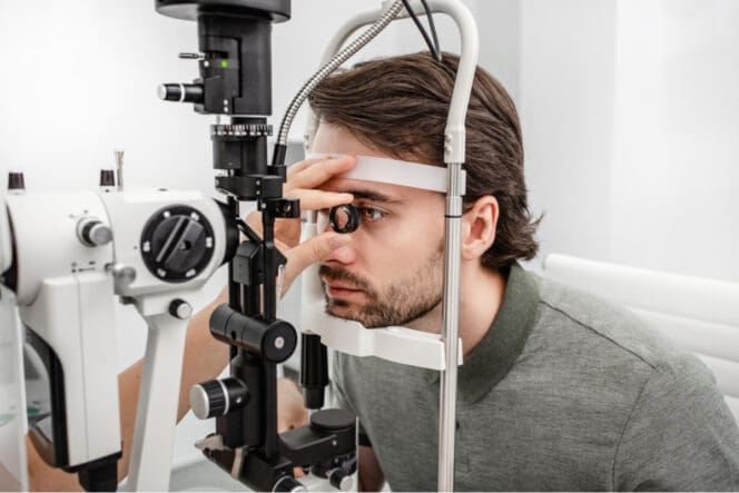
Eye doctors can diagnose retinoschisis during an eye examination of the back of the eye. Upon a detailed inspection with Optical Coherence Tomography (OCT), doctors can see any rips, tears and splits to the retina.
Other tests that a doctor may use to make a diagnosis are an electroretinogram (ERG) and ultrasounds.
Most retinoschisis cases do not affect your central vision and do not always require treatment. In fact, there is no current treatment regimen for degenerative retinoschisis. However, for certain complications, doctors may opt for vitrectomy surgery.
Children with hereditary retinoschisis may experience bleeding within their eye. This symptom requires treatment, and doctors advise to keep the eye as still as possible to promote coagulation. Once the bleeding stops, doctors turn to cryotherapy or laser treatments to close up damaged areas of the retina.
People with retinoschisis can also benefit from corrective glasses. Prescription eyewear can improve your quality of vision while also helping with myopia or hyperopia. Although they are useful in these cases, glasses will not correct or reverse nerve tissue damage resulting from retinoschisis.
Recently, there have been more investigative therapy studies to help with retinoschisis. These include the use of vitamin A, which is well known for its benefits on other genetic retinal diseases.
However, vitamin A appears to have no effect on retinoschisis as it is quite different from eye conditions such as retinitis pigmentosa, which benefit from the vitamin. More research is needed to understand the underlying causes leading to retinoschisis and possible treatment options.
Complications Linked to Retinoschisis
The most significant complication linked to retinoschisis is retinal detachment. The retina’s outer layer is anchored to the eye’s wall. When damage occurs to this anchor, it may lead to retinal detachment. While retinal detachment can happen to anyone, it is more prevalent in people who have retinoschisis.
When identified early enough, retinal detachments can be treated effectively. Positive outcomes from early diagnoses accentuate the importance of regular eye exams with your optometrist or ophthalmologist, especially if your family has a history of retinoschisis or other vision complications.
FAQs
Can retinoschisis be treated?
There are no treatment options available for retinoschisis today, although corrective eyeglasses can improve your vision. Children with hereditary retinoschisis who experience eye-bleeding can get help through cryosurgery or laser therapy.
How does retinoschisis occur?
Retinoschisis develops when the retina splits into two, which then affects your vision. The condition can either be hereditary or caused by old age. If you suspect you may have retinoschisis, you should talk to your eye specialist for further tests and a course of action.
What is the difference between retinoschisis and retinal detachment?
Retinal detachment normally collapses under the sclera depression area while retinoschisis moves according to the retinal area being depressed. In addition, retinoschisis is typically transparent and clear, meaning doctors can easily see the any rips, tears and other damage that has occurred.
Can you drive with retinoschisis?
Since retinoschisis damages the macula, it can interfere with your ability to make out certain colors and shapes in front of you. These problems with central vision can make certain tasks difficult, including driving and reading. Always check with your doctor if you can perform various tasks, like driving if you have retinoschisis.
References
-
Common Causes of Vision Loss in Elderly Patients. (July 1999). American Academy of Family Physicians.
-
X-linked Retinoschisis: A Clinical and Molecular Genetic Review. (March-April 2004) National Center for Biotechnology Information.
-
What Is a Detached Retina. (September 2021). American Academy of Ophthalmology.
-
X-linked Retinoschisis: Clinical Phenotype and RS1 Genotype in 86 UK Patients. (April 2005). Journal for Medical Genetics.
-
Laser Therapy in the Treatment of Diabetic Retinopathy and Diabetic Macular Edema. (September 2021). National Center for Biotechnology Information.
-
Vitamin A Derivatives as Treatment Options for Retinal Degenerative Diseases. (July 2013). National Center for Biotechnology Information.
-
Retinal Detachment Due to an Outer Retinal Tear Following Laser Prophylaxis for Retinoschisis. (December 2008). Journal of the American Medical Association.
Last Updated April 4, 2022
Note: This page should not serve as a substitute for professional medical advice from a doctor or specialist. Please review our about page for more information.