Human eyes function when the three parts of the eye work together to let in light and focus the light to the retina, which sends electrical signals through the optic nerve to the brain.
Eyes have three distinction sections — front, center and back. The front handles light input and focus. The center ensures the eye shape and structure holds steady. And the rear transmits signals to the brain about what is being looked at.
Vision issues and sight loss can occur in any of the three sections and with any parts of them.
Intro
The eyes are the second most complex organs in the human body after the brain. They contain many distinct structures to help you focus the light from your environment and turn it into useful visual information.
Your eyes cannot function properly without help from your brain. The tight link between the two organs means that sight is not a completely objective sense. The brain can distort or fill in visual information based on contextual cues and past experiences. This helps you to navigate the world around you even when your vision is limited.
Anatomy of the Eye
The human eye has many distinct parts that work together to help it perform its functions. The best way to understand these parts is to break them into three sections:
- Front
- Center
- Back
The Front
The front of the eye is the most complex part of the organ. It is where many of the structures that are used to focus light and produce clear vision.
Parts of the eye front include:
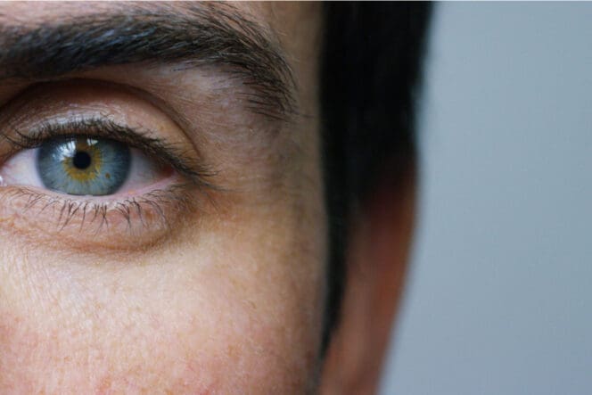
- The cornea: the clear layer on the outside of the eye which focuses light into the internal structures. It sits over the iris and pupil but does not extend over the sclera.
- The aqueous humor: the clear watery substance that fills the area between the cornea and the iris.
- The sclera: the white part of the eye that can be seen around the iris. This part of the eye sits in front of the iris and forms the front portion of the outer layer of the eye, holding the organ together.
- The pupil: the dark circle visible in the middle of the iris. The pupil fluctuates in size depending on the amount of light it is exposed to.
- The iris: the colored ring around the pupil. The iris controls the pupil and allows it to shrink (constrict) and grow (dilate) in response to lighting conditions.
- The ciliary body: the muscular structure that sits behind the iris and controls the lens. The ciliary body’s movements allow the lens to focus light onto the retina.
- The lens: the transparent structure that sits behind the iris and is attached to the ciliary body. It focuses the light passing through the eye onto the retina.
The Center
The center of the eye is full of vitreous fluid, a clear, jelly-like substance. Vitreous fluid provides the internal pressure the eye needs to hold its shape.
Vitreous fluid is transparent and uncolored, properties needed to allow light to pass through it and make its way to the retina at the back of the eye. Vitreous fluid contains no blood vessels or other structures that could get in the way of light that is traveling through the eye.
Vitreous fluid plays no active role in the visual system. What it does is support many of the other parts in performing their functions.
The pressure it exerts on the outer structures of the eye ensures that the light passing through the eye always travels the same distance. This is critical to keeping your sight consistent and predictable.
The Back
The back of the eye contains the parts that connect your eyes to your brain. It also contains the structures that keep the organ supplied with blood.
The parts in this section include:
- The macula: the part of the retina that contains light-sensitive cells. Macular cells help us see fine details when our eyes are focused on an object.
- The fovea: a small hollow in the center of the macula that concentrates light and sharpens your vision.
- The choroid: the layer that contains the blood vessels that feed the back of the eye. It is attached to the ciliary body and covers the rear two-thirds of the eye.
- The retina: the layer of nerves at the back of the eye. The retina contains special cells called photoreceptors (rods and cones). These cells convert the light that has passed through your eyes into electrical signals that can be understood by your brain.
- The optic nerve: the nerve that transmits visual information (now in the form of electrical impulses) from the eye to the brain.
How the Eyes Work
Every time you look at something, your eyes take those sights to the brain by following the same sight process:
- Light is reflected off the object you are looking at.
- The light enters your eyes through the cornea and is directed onto the pupil. If there is a lot of light present, the pupil contracts to reduce the amount of light that can get in. This reduces the amount of visual stimulus you must process. If there is little light, the pupil dilates to let in more light. This helps you get as much visual information about your environment as possible.
- Your lens and cornea work like a set of prisms to refract the light that is coming in. They bend it into a more concentrated form that brings the objects in front of you into focus.
- The light passes through your eye and eventually reaches your retina. The retina then transforms the light signals into electrical signals that your brain can understand.
- These signals are carried by your optic nerve until they reach the visual cortex at the back of your brain.
- Your brain interprets the electrical signals and forms an image of the environment you are in. Because of how light is reflected across refractive surfaces, any images in the light that hits your retina are upside down at first. Your brain automatically flips the image to help you better navigate the world around you.
If it is not possible for your body to carry out any of these steps, you will not be able to see correctly.
It is possible to have eyes that are anatomically capable of vision and still have no sight in those eyes. This can happen due to congenital brain abnormalities, brain damage or abnormal development.
Some studies have shown that it may be possible to regrow some of the visual pathways in the brain if they are lost. However, these findings have not yet been used to create a successful treatment for vision loss.
Today, sight lost because of a brain problem cannot be restored.
The Eye-Brain Connection
The connection between your eyes and brain has significant implications for two other domains related to vision: perception and prediction.
Perception
The eyes and the brain depend on each other to make sight possible. This connection means that vision is not an objective sense. When you look at an object, you do not necessarily see what it really is. Rather, you see what your brain interprets it to be.
Optical illusions are an example of this phenomenon. These images are carefully crafted to trick your brain into thinking it is seeing something that is not actually there. Many people have difficulty perceiving these images objectively even when they know the trick behind the illusion. They cannot alter their brain’s interpretation of the image, so they cannot see the real image.
Prediction
The eye-brain connection also helps you to better interpret your environment in poorly lit, busy, or cluttered conditions. When your eyes do not receive enough visual information to form a clear picture of your surroundings, your brain may fill in the gap with predictions based on context and what it has seen before.
The eye’s predictive function helps you to draw meaningful conclusions about your environment. For instance, if you see the front wheel of a tricycle peeking out from behind a nearby car, your brain quickly concludes that there is likely an entire tricycle behind the car. Your eyes do not have to see the tricycle for you to know it is there.
This information can help you take appropriate action, such as slowing down your car to make sure you can stop in time if a child rides the tricycle out in front of you.
References
-
Eye Anatomy and Function. (August 2020). MyHealth Alberta.
-
Anatomy of the Eye. (2022). Kellogg Eye Center: University of Michigan Medicine.
-
How the Eyes Work. (March 2022). National Eye Institute.
-
Use it or lose it: Visual activity regenerates neural connections between eye and brain. (July 2016). National Institutes of Health.
-
Neuro-Visual Disorders. (2022). Johns Hopkins Medicine.
-
Reconnecting Eye to Brain. The Journal of Neuroscience.
-
Eyes. (September 2021). Cleveland Clinic.
-
How the brain decides what we see. (January 2005). Journal of the Royal Society of Medicine.
-
Visual illusions and neurobiology. (December 2001). Nature Reviews Neuroscience.
-
Anatomy of the eye. (November 2017). Moorfield Eye Hospital: National Health Service.
-
10 fun facts about eyes. (July 2016). Washington State University: Cougar Health Services.
Last Updated April 19, 2022
Note: This page should not serve as a substitute for professional medical advice from a doctor or specialist. Please review our about page for more information.